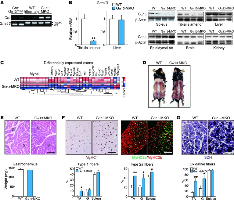
Figure 2. Loss of Gα13 causes a skeletal muscle switch to the oxidative phenotype.
(A) PCR analysis using tail genomic DNA from WT C57BL/6 mice, WT littermates (Gna13fl/fl), and Gα13-MKO (Ckmm-Cre+ Gna13fl/fl) mice. (B) qPCR analyses of 3 samples each and immunoblots for Gα13 in the indicated tissues. Twelve-week-old mice were fasted overnight prior to sacrifice. (C) Global gene expression analysis. RNA was isolated from soleus muscles from mice of the indicated genotypes and hybridized to Affymetrix exon arrays. (D) Dorsal view of skinned WT and Gα13-MKO mice 35 weeks after birth. Enhanced larger images are shown in Supplemental Figure 1B. (E) H&E staining and wet weight of hind limb muscles. G, gastrocnemius; S, soleus. (F) Representative immunohistochemical images of tibialis anterior muscles from 12-week-old WT or Gα13-MKO mice using specific antibodies for each of the fiber types (n = 3). Myofiber types in the indicated tissues were quantified. (G) Representative histochemical staining of SDH enzymatic activity in tibialis anterior muscles (n = 3–4 per genotype). Myofibers with high SDH activity were quantified. Scale bars: 200 μm (E–G). For B and E–G, data represent the mean ± SEM. *P < 0.05 and **P < 0.01, by Student’s t test. G, gastrocnemius.

