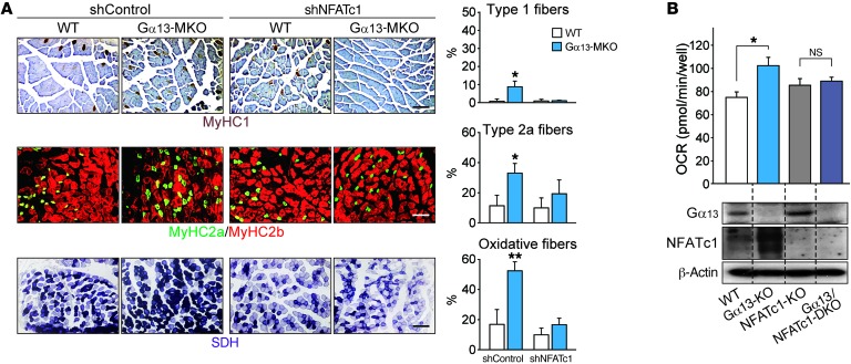
Figure 5. NFATc1 mediates the oxidative conversion of myofibers by Gα13 ablation.
(A) Immunostain images for myosin heavy chains and histochemical assays for SDH activity of tibialis anterior muscles 14 days after electroporation-mediated gene delivery. Each mouse of the indicated genotype received a control shRNA vector in 1 limb and a plasmid expressing shNFATc1 in the contralateral limb. Type 1 and 2a myofibers and those with high SDH activity were quantified. Scale bars: 200 μm. (B) Respiration assay and immunoblots. Basal OCRs were determined in C2C12 myotubes of the indicated genotypes, which were prepared by CRISPR-mediated gene editing (n = 3 each). Blots were obtained from samples run on parallel gels. DKO, double-KO. All data represent the mean ± SEM. *P < 0.05 and **P < 0.01, by Student’s t test.

