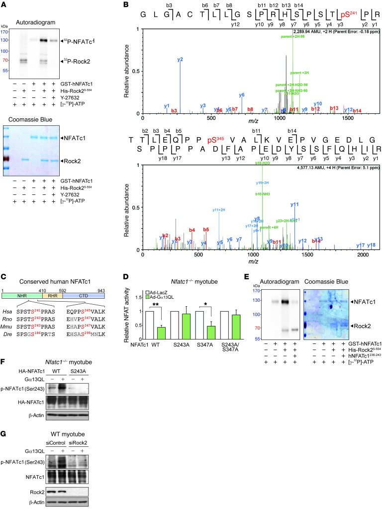
Figure 7. Rock2 phosphorylates NFATc1 at Ser243.
(A) In vitro kinase assay. Recombinant proteins and [γ-32P]-ATP were incubated with or without Y-27632 (10 μM), and phosphorylation was visualized by autoradiography. (B) LC-MS/MS analysis of human NFATc1 after incubation with human Rock25-554. (C) Schematic illustration of NFATc1 protein domains and sequence comparisons of positive hits among different species. (D) NFATc1 transcription activity assay. NFATc1-KO C2C12 myotubes were transfected with an NFATc1 reporter plasmid in combination with a WT or serine-to-alanine mutant NFATc1 plasmid (n = 3 each). Data represent the mean ± SEM. *P < 0.05 and **P < 0.01, by Student’s t test. (E) In vitro kinase assay. An 8-mer peptide (hNFATc1236-243) surrounding the Ser241 residue was added to the reaction mixture where indicated. Kinase activity was determined by incorporation of the [γ-32P]-ATP label. (F and G) Immunoblots for phosphorylated NFATc1 (p-NFATc1) (Ser243). (F) NFATc1-KO C2C12 myotubes were transfected with a plasmid encoding WT NFATc1 or its S243A mutant. Adenoviral infection of Gα13QL was done 12 hours after the transfection, and samples were prepared 24 hours later. (G) C2C12 myotubes were transfected with siRNA against Rock2 or with control siRNA. After 12 hours, Gα13QL was transfected via adenovirus, and the samples were prepared 12 hours later.

