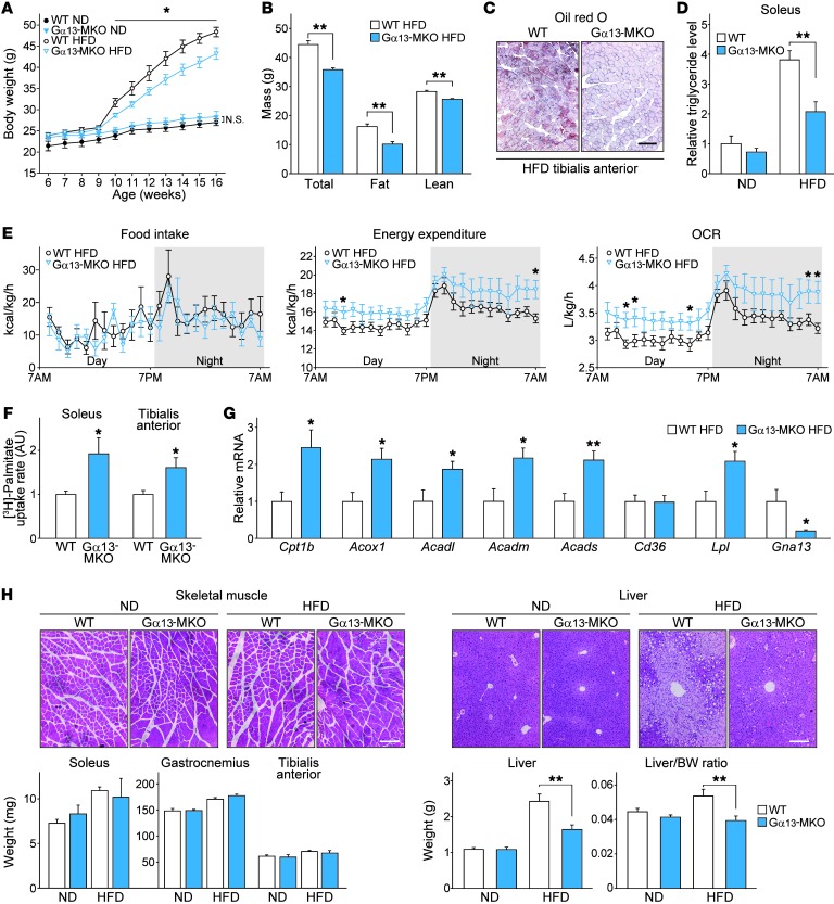
Figure 9. Gα13-MKO protects mice from diet-induced adiposity with increased fatty acid metabolism.
(A–H) Nine-week-old WT or Gα13-MKO mice were fed a ND or a HFD. After 9 weeks of HFD feeding, the mice were fasted overnight and then sacrificed. (A) Body weight gains (n = 6–8 each). (B) Determination of fat and lean mass in HFD-fed WT and Gα13-MKO mice using nuclear magnetic resonance (n = 8 each). (C) Oil red O staining of tibialis anterior after HFD feeding. Scale bar: 200 μm. (D) Relative triglyceride levels in mouse soleus muscles normalized with protein levels (n = 6–8 each). (E) In vivo energy balance. Food intake, energy expenditure, and OCR were analyzed in mice housed in individual metabolic cages (n = 8 each). (F) Ex vivo fatty acid uptake assay (n = 3 each). Muscles were freshly isolated from mice of each genotype, incubated in a medium containing [3H]-palmitate acid for 30 minutes, and lysed for scintillation counting. (G) qPCR assays for transcripts of the genes associated with lipid uptake and oxidation (n = 6–8 each). (H) H&E-stained images of skeletal muscles and liver and data on skeletal muscle and liver weights and liver/body weight ratios (n = 6–8 each). Scale bars: 200 μm. For A, B, and D–H, data represent the mean ± SEM. *P < 0.05 and **P < 0.01, by Student’s t test.

