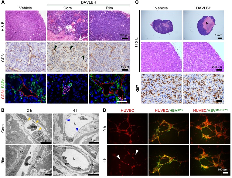
Figure 1. Vessels in the tumor periphery with higher pericyte coverage show resistance to DAVLBH.
For in vivo experiments, mice bearing MDA-MB-231 xenografts received i.v. injection of 2 μmol/kg DAVLBH, and the tumors were harvested 2 hours, 4 hours, or 2 days later. (A) H&E, CD31, and FAPα/CD31 staining show a marked vascular disruption in tumor core, but not in peripheral tumor, after 4 hours of treatment (n = 5). The black arrowheads indicate the loss of ECs, and autofluorescent rbc appear as pink points in FABα/CD31-stained sections. Top row scale bars: 200 μm. Middle row scale bars: 50 μm. Bottom row scale bars: 50 μm. (B) The transmission electron microscope images show the effects of DAVLBH on tumor vessels. The yellow arrowheads indicate EC blebbing, and the blue arrowheads indicate the loss of ECs. L, lumen; PC, pericyte. Top row scale bars: 5 μm. Bottom row left scale bar: 2 μm. Bottom row right scale bar: 10 μm. (C) H&E staining shows extensive necrosis Top row scale bars: 1 mm. Middle row scale bars: 200 μm. Bottom row scale bars: 50 μm. (N) in tumor core after 2 days of treatment; Ki67 staining shows similar proliferation between 2 groups (n = 5). (D) The EC- (red) and pericyte-cocultured (green) systems show DAVLBH selectively damages the HUVEC tubes (white arrows), but has negligible effect on the HBVPNC-HUVEC and HBVPFAPα-WT-HUVEC–cocultured tubes (n = 3). Scale bars: 100 μm.

