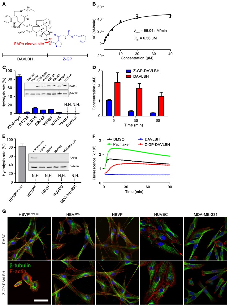
Figure 2. Z-GP-DAVLBH is selectively hydrolyzed by FAPα to release DAVLBH to disrupt the cytoskeleton of FAPα-expressing pericytes.
(A) Structure of Z-GP-DAVLBH. (B) Evaluation of the enzymatic kinetics of rhFAPα on Z-GP-DAVLBH. The substrate-velocity curve for cleavage of Z-GP-DAVLBH by rhFAPα (5 ng/ml) is shown (n = 3). (C) Enzymatic efficacy of engineered FAPα-expressing cells on Z-GP-DAVLBH. HEK-293T cells were transiently transfected with vector, WT FAPα, or mutant FAPα plasmids (R123A, E203A, E204A, Y656F, N704A). Z-GP-DAVLBH (10 μM) was cocultured with cells at 37°C for 2 hours, and hydrolysis was analyzed by LC/MS. Quantification of the hydrolysis rate is shown (n = 3). N.H., no hydrolysis. (D) Evaluation of the hydrolysis for Z-GP-DAVLBH in MDA-MB-231 tumor xenografts (n = 5). The concentrations of Z-GP-DAVLBH and DAVLBH in tumors were detected at 5, 30, and 60 minutes after i.v. injection of Z-GP-DAVLBH (2.0 μmol/kg). (E) Enzymatic ability of HBVPFAPα-WT, HBVPNC, HBVPs, HUVECs, and MDA-MB-231 against Z-GP-DAVLBH. Quantification of the hydrolysis rate is shown (n = 3). (F) Inhibition of tubulin polymerization by DAVLBH and Z-GP-DAVLBH in vitro. Purified porcine brain tubulin was incubated with the tested compounds at 1 μM. Effects on tubulin polymerization were monitored by fluorescence value measurement, with excitation at 360 nm and emission at 420 nm every 1 minute for 90 minutes at 37°C. Paclitaxel (3 μM) was used as positive control agent. (G) The effect of Z-GP-DAVLBH on the β-tubulin cytoskeleton of HBVPFAPα-WT, HBVPNC, HBVPs, HUVECs, and MDA-MB-231 cells (n = 3). The cells were treated with Z-GP-DAVLBH (2.5 nM) for 30 minutes, and β-tubulin (green) and F-actin (red) were observed with a confocal microscope. Data are shown as mean ± SEM. Scale bar: 50 μm.

