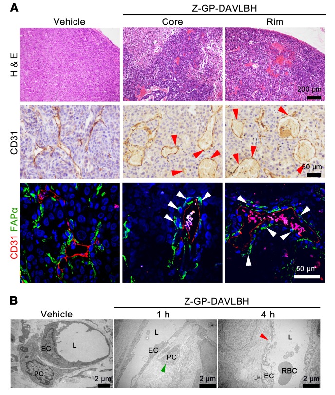
Figure 4. Z-GP-DAVLBH targeting pericytes disrupts vessels in both the tumor core and periphery.
Tumors were harvested at 1 hour or 4 hours after MDA-MB-231 xenografts received i.v. injection of 2.0 μmol/kg Z-GP-DAVLBH (n = 5). (A) H&E staining shows vascular disruption in both the tumor core and the periphery; CD31 staining shows “hole-like” disruptions in the vessels (red arrowheads); FAPα/CD31 staining shows the shrinkage and detachment of pericytes (white arrowheads). Tumors were harvested after 4 hours of treatment. Top row scale bars: 200 μm. Middle row scale bars: 50 μm. Bottom row scale bars: 50 μm. (B) The transmission electron microscope images show the effects of Z-GP-DAVLBH on vessels in the tumor periphery. The green arrowhead indicates the shrinkage and detachment of tumor pericyte; the red arrowhead indicates a “gap” disruption between 2 ECs. Scale bars: 2 μm

