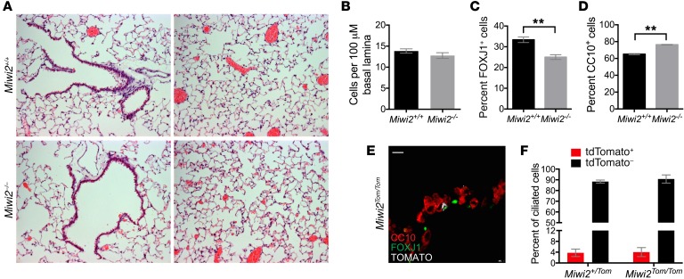
Figure 5. MIWI2 regulates pulmonary epithelial cell composition.
(A) H&E staining of lung sections from uninfected Miwi2+/+ and Miwi2–/– mice. (B–D) Lung sections from uninfected Miwi2+/+ and Miwi2–/– mice were immunostained for CC10, FOXJ1, and DAPI, and the total numbers of airway cells (B), ciliated cells (C), and club cells (D) were quantified. n = 4, 3 mice per group, data presented as mean ± SEM. **P < 0.01 as determined by unpaired t test. (E) Lung sections from Miwi2Tom/Tom mice immunostained for red fluorescent protein (RFP, white), FOXJ1 (green), and CC10 (red). Scale bars: 10 μm. (F) Flow cytometric quantification of tdTomato-positive and tdTomato-negative cells as a percentage of live, CD45–EpCAM+CD24hi ciliated cells; n = 3, 4 mice per group, data presented as mean ± SEM.

