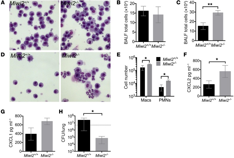
Figure 6. MIWI2 regulates pulmonary innate immunity during pneumonia.
(A and B) BALF cell cytospins (A) and total BALF cell counts (B) of uninfected Miwi2+/+ and Miwi2–/– mice. n = 4, 4 mice per group, data are expressed as mean ± SEM. Microscopic images were obtained using the ×40 objective. (C–G) Total BALF cell counts (C), BALF cytospins (D), BALF differential counts (E), BALF CXCL2 protein (F), and BALF CXCL1 protein (G) were measured from Miwi2+/+ and Miwi2–/– mice infected i.t. with S. pneumoniae for 4 hours. n = 6, 7 mice per group, data are expressed as mean ± SEM. *P < 0.05, **P < 0.01 as determined by unpaired t test. Microscopic images were obtained using the ×40 objective. (H) Lung CFU was determined in Miwi2+/+ and Miwi2–/– mice infected i.t. with S. pneumoniae and harvested 30 hours later. n = 6, 9 mice per group, data represented as mean ± SEM. *P < 0.05 as determined by unpaired t test. Dotted line indicates input CFU.

