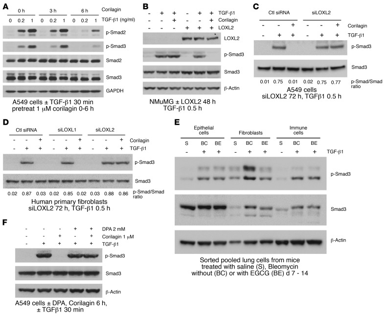
Figure 4. Corilagin inhibition of TGF-β1 signaling is dependent on LOXL2 activity.
(A) A549 cells pretreated with 1 μM corilagin for 0, 3, or 6 hours were stimulated with different doses of TGF-β1 (0, 0.2, or 1 ng/ml) for 30 minutes, and the cell lysates were blotted for p-Smad2, p-Smad3, Smad2, Smad3, and β-actin. (B) NMuMG cells transfected with human LOXL2 or empty vector were pretreated with 1 μM corilagin or DMSO for 6 hours and then incubated without or with TGF-β1 for 30 minutes. The cell lysates were blotted for LOXL2, p-Smad3, Smad3, and β-actin. (C and D) A549 cells transfected with siRNA to LOXL2 (C) and primary human lung fibroblasts transfected with siRNAs to LOXL1 or LOXL2 (D) were pretreated with 1 μM corilagin or DMSO for 6 hours before incubation without or with TGF-β1 for 30 minutes. The cell lysates were blotted for p-Smad3 and Smad3. The ratio of p-Smad3/Smad3 for each lane is shown. (E) Mouse lung epithelial cells, fibroblasts, and immune cells sorted from mice treated for 14 days with saline (S), bleomycin with vehicle control (BC), or bleomycin with 7 days oral EGCG (100 mg/kg) (BE) were immediately treated with TGF-β1 for 30 minutes and cell lysates blotted for p-Smad3, total Smad3, and β-actin. n = 5 for each group. (F) A549 cells were pretreated with 1 μM corilagin with or without 2 mM DPA for 6 hours before TGF-β1 stimulation for 30 minutes. The cell lysates were blotted for p-Smad3, Smad3, and β-actin. The data from A–D and F are representative of at least 3 experiments with similar results.

