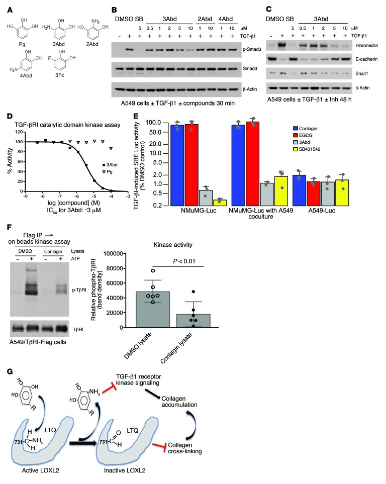
Figure 6. Generation of a novel nondiffusible TβRI kinase inhibitor.
(A) Structure of pyrogallol (Pg), 3Abd, and derivatives. (B) A549 cells were stimulated with TGF-β1 for 30 minutes and lysates immunoblotted for p-Smad3, Smad3, and β-actin. Treatment without preincubation: 3Abd (0.5–10 μM); 2Abd or 4Abd (1, 10 μM). (C) A549 cells were stimulated with TGF-β1 for 48 hours and lysates immunoblotted for LOXL2, fibronectin, E-cadherin, Snail1, and β-actin. 3Abd was added at concentraions ranging from 0.5 to 10 μM. (D) Purified ALK5/TβRI catalytic domain kinase assay was performed with 10 doses of 3Abd or Pg starting from 100 μM. Kinase activity was indicated by 33P-ATP signals, and IC50 of 3Abd was calculated as approximately 3 μM. B–D are representative of 3 experiments with similar results. (E) SMAD-binding element (SBE) reporter–transfected NMuMG and A549 cells were seeded into a 96-well plate. Cocultured wells were seeded with 5,000 transfected NMuMG cells and 25,000 nontransfected A549 cells. Cells were pretreated with or without 1 μM corilagin, 1 μM EGCG, 10 μM 3Abd, or 5 μM SB431542 for 6 hours and stimulated with TGF-β1 overnight before lysis for luciferase assay. Data are presented as percent TGF-β1–induced SBE luciferase activity of DMSO control in log scale. Mean ± SD, n = 3. (F) Flag-tagged TβRI and TβRII were immunoprecipitated from A549 cells and in vitro kinase assay performed on beads exposed to lysate pretreated with corilagin or DMSO. The final reaction was eluted and analyzed by immunoblotting for phosphotyrosine and TβRI. The phosphotyrosine bands were quantified using ImageJ and normalized to DMSO control. Data represent mean ± SD. P value by unpaired 2-tailed t test of 6 separate experiments. (G) Schematic overview of mechanism. A trihydroxyphenolic-containing compound engages active LOXL2, initiating auto-oxidation of K731 and creating a key allysine inactivating the enzyme. In the process, a 3Abd-like metabolite is generated that then blocks TβRI kinase. The combined effects block pathological collagen accumulation.

