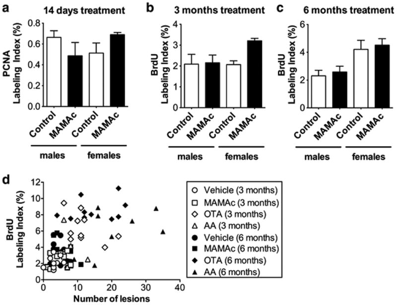
Figure 5.

(a-c) Assessment of MAMAc effects on proliferation via (a) proliferating-cell-nuclear-antigen (PCNA) S-phase labeling indices (LI %) in the renal cortex of 14-days MAMAc treated male and female Eker rats (n=3), or via (b, c) BrdU S-phase labeling indices of male and female Eker rats treated with MAMAc for 3 (b) and 6 months (c), respectively (n=5). (d) Pearson correlation analysis of BrdU labeling indices versus the total number of renal lesions determined by renal histopathology in rats (N = 5 per group), either 3 months (white) or 6 months (black) treated with vehicle (circles), MAMAc (squares), ochratoxin A (OTA, diamonds), and aristolochic acid (AA, triangles). Data represent means ± SEM. One-way ANOVA with Tukey's post-hoc test (a-c) was used to test for statistical significance.
