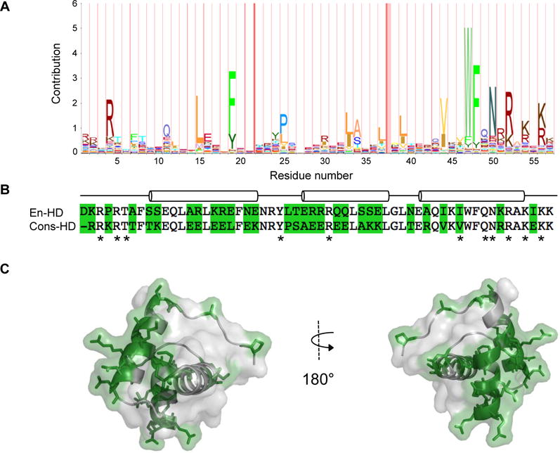
Figure 1. Sequence conservation and consensus of the homeodomain family.

A) The sequence logo, where residue height is proportional to degree of conservation, is calculated from the Pfam version 26.0 homeodomain seed sequence from 12/21/2012. B) Sequence alignment of engrailed- and consensus-HDs. Sequence differences are shaded green. Residues that contact DNA are indicated with asterisks. The positions of the three helices in the engrailed-HD are indicated with cylinders. C) Sequence differences (shaded in green with side chains shown) mapped onto the crystal structure of engrailed-HD (PDB: 1ENH).
