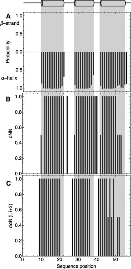
Figure 3. Secondary-structure of consensus-HD determined by NMR spectroscopy.

A) Plot of chemical shift-based secondary structure predictions by TALOS+29 B) 1HNi- 1HNi+1 and C) 1Hαi- 1HNi+3 NOEs. Strong NOEs were given value of one. Weak but detectable NOEs were given a value of 0.5. Positions with no bar showed no detectable NOE. Helix boundaries (top) are based on the engrailed-HD structure. Sequence positions are as in alignment in Figure 1B where position 1 corresponds to the N-terminal D in engrailed-HD and position 2 corresponds to the N-terminal R in consensus-HD. Conditions: 25 mM sodium phosphate, 150 mM NaCl, pH 7, 20 °C.
