Figure 2.
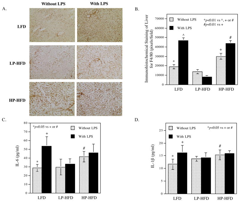
The effect of LPS and diets on hepatic and systemic inflammation.
(A and B) After mice were treated with LPS or vehicle PBS in combination with LFD, LP-HFD, or HP-HFD, livers were dissected and subjected to immunohistochemical staining of F4/80 to detect macrophages. Representative photomicrographs of hepatic tissue sections with F4/80 immunostaining for all 6 groups (A) and quantification of F4/80-positive staining area (B) were shown. C and D. Serum IL-6 (C) and IL-1β (D) were quantified at the end of the study. The data presented are mean ± SD (n=6–9).

