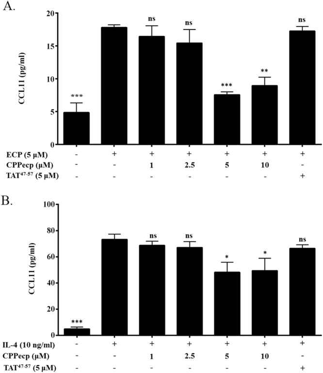
Figure 2.

(A) CCL11 secretion in CPPecp and ECP co-treated BEAS-2B cells. BEAS-2B cells were starved with serum free medium for 24 h, followed by stimulation with ECP. Cells were stimulated with PBS as negative control and ECP in the absence of CPPecp or presence of 1, 2.5, 5, and 10 μM CPPecp at 37 °C for 24 h. Cells were stimulated with ECP in the presence of 5 μM TAT. CCL11 protein level was determined by ELISA kit employing CCL11 antibody. The data represented mean ± SD of at least three independent experiments. ***p < 0.001; **p < 0.01. (B) CCL11 expression in CPPecp and IL-4 co-treated BEAS-2B cells. BEAS-2B cells were starved with serum free medium for 24 h, followed by stimulation with PBS or 10 ng/ml IL-4. Cells were stimulated with 10 ng/ml IL-4 in the absence or presence of 1, 2.5, 5 and 10 μM CPPecp at 37 °C for 24 h. Cells were stimulated with IL-4 in the presence of 5 μM TAT. CCL11 protein level was determined by ELISA kit employing CCL11 antibody. The data represented mean ± SD of at least three independent experiments. ***p < 0.001. *p < 0.05.
