Figure 1.

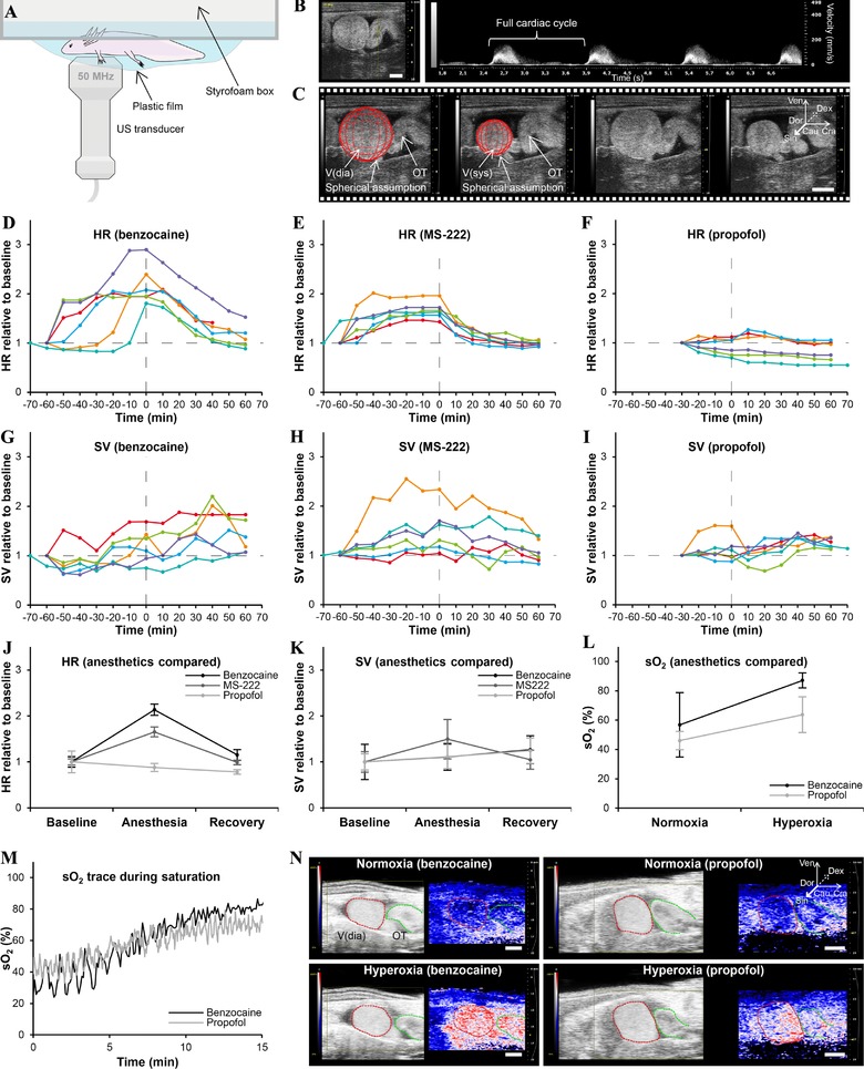
Effect of benzocaine, MS‐222, and propofol on cardiovascular function. (A) Experimental TTE setup. (B) Pulsed wave velocity TTE allows for HR measurements (panel to the left shows image probe position). (C) Brightness mode TTE allows for SV measurements assuming a spherical shape of the ventricle. (D)−(F) HR plotted over time (0 h is at full anesthesia) for all animals for benzocaine (D), MS‐222 (E), and propofol (F). (G)−(I) SV plotted over time for all animals for benzocaine (G), MS‐222 (H), and propofol (H). (J)−(K) Comparison of mean HR (J) and SV (K) for the three anesthetics at baseline, full anesthesia, and full recovery. (L) Mean intracardiac blood oxygen saturation (sO2) at normoxia and 100% ambient oxygen saturation for benzocaine and propofol. (M), (N) Representative sO2 traces (M) and sO2 images (N) during ambient oxygen saturation for benzocaine and propofol. V(dia), ventricle in diastole; V(sys), ventricle in systole; Ven, ventral; Dor, dorsal; Cau, caudal; Cra, cranial; Dex, right; Sin, left
