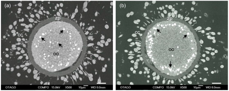
Figure 3.
SEM images of lipid distribution in mature sheep oocytes (oo) surrounded by zona pellucida (zp) and expanded cumulus cells (cc). Lipid droplets (arrows) are either distributed: throughout the cytoplasm (a); or in a peripheral location (b). A higher proportion of oocytes from adult sheep had evenly distributed lipid droplets than those from prepubertal sheep that had predominantly peripherally located droplets. Scale bar = 20 μm.

