
Figure 1.
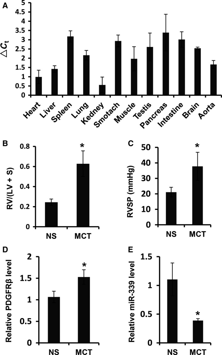
MiR‐339 expression was decreased in pulmonary arteries of MCT‐induced PAH rats. (A) Histogram showed the miR‐339 expression in various organs in rats, ▵Ct = Ct(miR‐339)‐Ct(sno202), n = 5. (B) The ratio of right ventricle (RV) and left ventricle (LV) plus ventricular septum (S) RV/(LV+S) in MCT‐induced rats was elevated significantly (n = 4) compared with that in the controls (n = 4). (C) Right ventricular systolic pressure (RVSP) in MCT‐induced rats was elevated significantly (n = 4) compared with that in the controls (n = 4). (D) Expression of PDGFR β mRNA in pulmonary arteries of MCT‐induced rats was increased significantly (n = 4) compared with that in the controls (n = 4). (E) Expression of miR‐339 in pulmonary arteries of MCT‐induced rats was decreased significantly (n = 4) compared with that in the controls (n = 4). MCT represented MCT‐treated rats, NS represented normal saline‐treated rats; different significance was assessed with Student's t test, *P < 0.01.