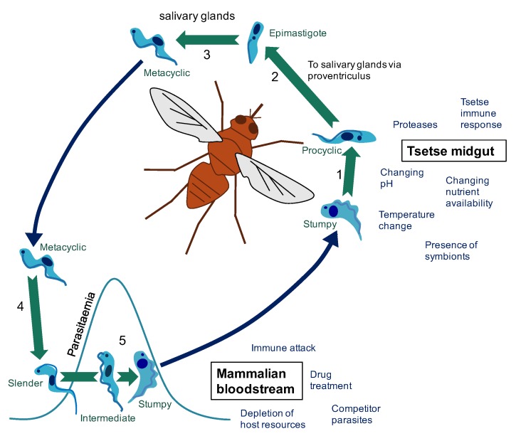
Figure 1.
The changing environments of the T. brucei life cycle. (1) Following a tsetse fly blood meal on an infected mammal (blue arrow), T. brucei stumpy cells reaching the tsetse midgut differentiate to procyclic cells. (2) Development of T. brucei proceeds through the tsetse midgut to the proventriculus. (3) Differentiation to epimastigote cells is followed by production of mammalian-infective metacyclic cells in the salivary gland. (4) On transfer to a new mammalian host via a tsetse fly bite (blue arrow), metacyclic cells differentiate to bloodstream forms. (5) In the bloodstream, proliferative slender cells elevate the parasitaemia until accumulation of a density-dependent signal triggers differentiation to the cell-cycle arrested stumpy form (via an intermediate stage). The stumpy form of T. brucei faces a particular challenge in that it must survive long enough in the bloodstream to be transmitted, but it must also survive long enough in the tsetse midgut to differentiate to the next life cycle stage and establish infection. These different environments pose different challenges that must be overcome to enable the continuation of the parasite life cycle. For example, in the mammalian host (red section), parasites must evade immune attack to ensure parasite survival and avoid excessive exploitation of host resources to ensure the host survives long enough to enable successful transmission to the tsetse fly vector. On uptake to the tsetse fly midgut (orange section), the parasites face a shift in nutrient availability and temperature among other challenges. For a review of factors effecting establishment of tsetse fly midgut infection see [1].

