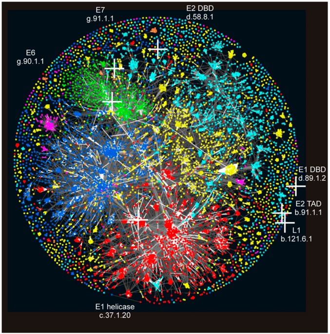
Figure 2.
Location of PV domains in the “Galaxy of folds”. PV structural domains are marked by white crosses and visualised on protein domain space. Domains in Structural Classification of Proteins (SCOP) were clustered using the software CLANS based on their all-against-all pairwise similarities, as measured by HHsearch p-values [34]. Domains are coloured according to their SCOP class: all-a (blue); all-b (cyan); a/b (red); a + b (yellow), small proteins (green); multi-domain proteins (orange); and membrane proteins (magenta). PV protein domain name and SCOP identifier are indicated.

