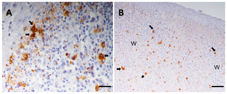
Figure 5.
Immunohistochemical detection of ASFV protein P30 on wax-embedded tissue sections from pigs inoculated with the highly virulent ASFV isolate OURT88/1 and euthanized at day 5 post-infection. (A) Tonsil, Bar 40 μm. Lymphoid follicle with severe lymphoid depletion. Observe the presence of infected macrophages (arrows) close to areas where lymphocytes show characteristic features of apoptosis such as reduced size and hyperchromatic nuclei. Cell debris and apoptotic bodies, many of them immunolabeled, are also observed; (B) spleen, Bar 80 μm. Note the presence of infected cells, mainly macrophages (arrows), along with pyknotic cells, cell debris and apoptotic bodies immunolabeled against P30 in white pulp areas (WP) with severe lymphoid depletion.

