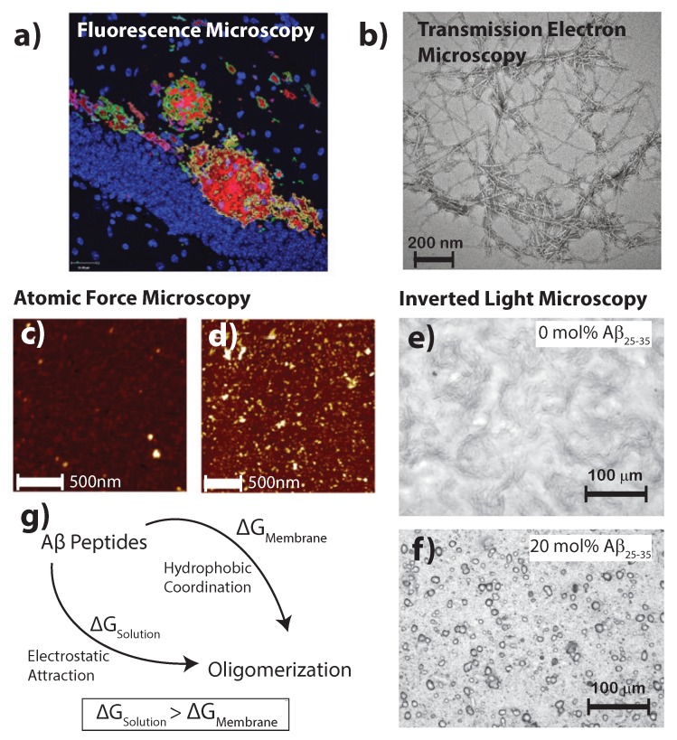
Figure 2.
(a) Fluorescent secondary antibodies to the A plaques in mouse models of Alzheimer’s disease are shown as per Fisher et al. [24] (copyright PLoS ONE, 2010); (b) Transmission electron microscopy of crystalized A photofibrils in solution from Chen et al. [25] (copyright PLoS ONE, 2012); Atomic force microscopy images in liquid shows in healthy (c) and diseased (d) membrane models; bars correspond to 500 and 1000 nm, respectively, and the image adapted from the preprint by Drolle et al. [19] (copyright arXiv, 2017); Optical microcopy images of (e) a pure 1-palmitoyl-2-oleoyl-sn-glycero-3-phosphocholine (POPC)/1,2-ditetradecanoyl-sn-glycero-3-phospho-L-serine (DMPS) membrane and (f) POPC/DMPS + 20 mol % A. While the pure lipid matrix shows a smooth surface, inclusions were observed at peptide concentrations of 10 and 20 mol % [21] (copyright Royal Society of Chemistry, 2016); (g) The consenses of literature suggests the formation of A aggregates is more favorable in the presence of a membrane than in pure solution.

