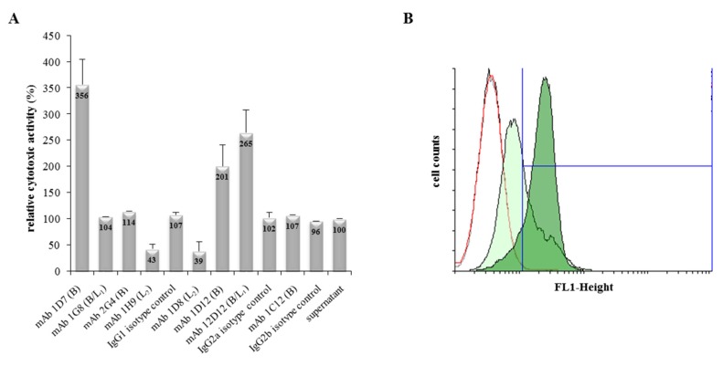
Figure 2.
Influence of mAbs on Hbl toxicity. (A) WST-1-Bioassay on Vero cells. Supernatant of F837/76 ∆nheABC was applied as serial dilution. mAb (10 μg/well) was added to each dilution and incubated with the cells for 24 h. Cytotoxicity titres were determined by addition of WST-1. The reciprocal titre of the untreated B. cereus supernatant was set to 100%. (B) Flow cytometry results of Vero cells treated with rHbl B. Cells were incubated for 1 h with either only buffer (black curve), Hbl B-specific mAb 1D7 (red curve), rHbl B (light green filled) or rHbl B + mAb 1D7 (dark green filled). Cell-bound rHbl B was detected by using Alexa Fluor® 488-labelled mAb 1G8 (Hbl B-specific).

