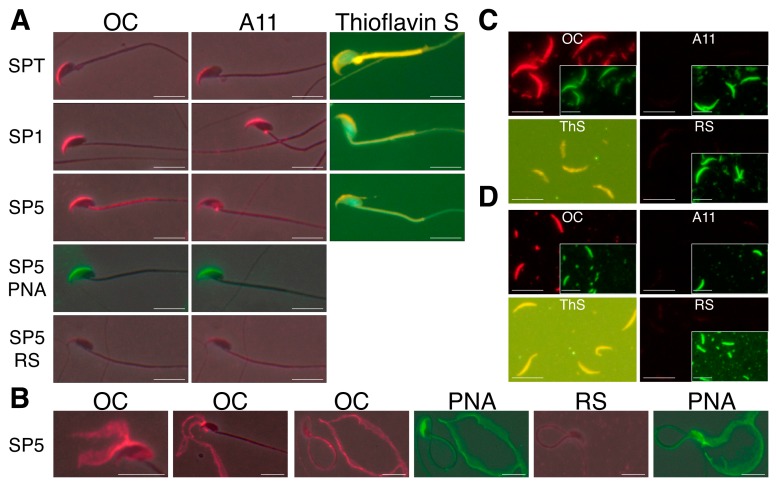
Figure 1.
Amyloids are present in mouse sperm acrosomes and isolated AM. Immunofluorescence analysis (IIF) using anti-fibrillar amyloid OC and anti-oligomeric amyloid A11 antiserum and thioflavin S staining showed the presence of amyloid in (A) intact acrosomes from testicular (SPT), caput epididymal (SP1), and cauda epididymal spermatozoa (SP5); (B) mechanically disrupted acrosomal shrouds from cauda epididymal spermatozoa; and isolated AM from (C) caput epididymal and (D) cauda epididymal spermatozoa. RS, normal rabbit serum. Staining with fluorescein isothiocyanate peanut agglutinin (FITC-PNA) was used as a marker for acrosomal material. Scale bar, 10 µm; insert, FITC-PNA staining shown at 40% reduction. Reprinted from [24].

