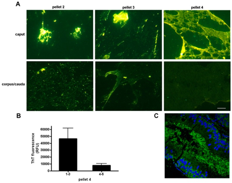
Figure 3.
Regional changes in thioflavin staining in the mouse epididymis. (A) Particulate material of varying molecular mass was obtained from the caput and corpus-cauda epididymal luminal fluid and stained with thioflavin S. Pellet 2, 5000× g; pellet 3, 15,000× g; pellet 4, 250,000× g. All images were captured with the same exposure times. Bar, 5 µm; (B) Thioflavin T fluorescence in pellet 4 obtained from the caput (regions 1–3) and corpus-cauda epididymis (regions 4–5) as determined in a plate assay. Mean + standard error of the mean (SEM) of three experiments; (C) Indirect immunofluorescence analysis of amyloid in the caput epididymal lumen using the anti-oligomeric amyloid antibody A11 (green fluorescence) (Muthusubramanian and Cornwall, unpublished observations). Blue, DAPI staining of epididymal epithelial cell nuclei. Modified and reprinted from [52].

