
Figure 8.
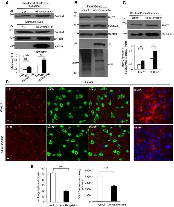
Overexpression of αB-crystallin rescues defective exosome secretion from KI astrocytes. A, Mouse αB-crystallin-HA plasmid was transfected into WT astrocytes. Flotillin-1 (Flotillin-1/GAPDH) remained unchanged in the astrocytic lysate (t = 0.203, df = 4, p = 0.1253 Student's t test, n = 3 independent cultures) but increased in exosomes (flotillin-1 in exosomes/in cell lysate) derived from αB-crystallin overexpressed in HD KI astrocytes compared with exosomes from astrocytes that received a control plasmid transfection (t = 2.985, df = 4, p = 0.0021, Student's t test, n = 3 independent cultures). B, C, Western blotting verified αB-crystallin-V5 expression in vivo. HSC70 and flotillin-1 increased in purified exosomes (Hsc70, t = 6.548, df = 4, p = 0.0028; flotillin-1, t = 4.503, df = 4, p = 0.0108; Student's t test, n = 3 αB-crystallin-injected mice), but not in cell lysates from the αB-crystallin-V5-injected striatum compared with an adenoviral-GFP control injection. Levels of mHtt aggregates decreased in the αB-crystallin-V5-injected region. D, High-magnification (63× objective) micrographs showing GFAP staining and mHtt aggregates (red), which are also shown in the merged images with NeuN (green) and nuclei stained by Hoechst (blue), in the striatum of 10-month-old HD mice. Scale bars, 5 μm. E, Quantitative analysis of the GFAP immunofluorescent density showing that GFAP staining and the number of mHtt aggregates significantly decreased in the αB-crystallin-V5-injected striatum compared with the control striatum (mHtt aggregates, control = 51.87 ± 0.53/image vs Ad-αB-crystallin = 19.28 ± 0.67/image, t = 38.13, df = 118, p = 3.42621E-68; GFAP fluorescence intensity, t = 20.46, df = 118, p = 1.30096E-40; Student's t test, n = 60 images in each group from 3 HD KI mice). *p < 0.05, **p < 0.01, ***p < 0.001.