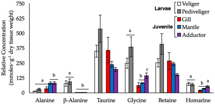
Figure 3.
The mean relative concentrations of alanine, β-alanine, taurine, glycine, betaine, and homarine (±SE) are shown for larval and juvenile mussels. Larval samples were analyzed at both the veliger (white bars; n = 3) and pediveliger (gray bars; n = 4) stages, while data from the gill (black bars), mantle (red bars), and adductor muscle (blue bars) were obtained from the tissues of individual juveniles (n = 5). Letters denote significant differences between the stages or tissues (at an experiment-wide α = 0.05).

