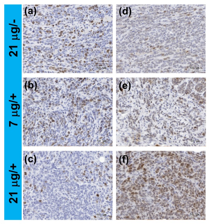
Figure 8.
In vivo analysis for a murine 4T1 breast cancer model based on 980-nm laser light-trigged photodynamic therapy (PDT) using the hybrid nanoparticles as the photosensitizer. The tumors were treated with lower (7 μg) and higher (21 μg) dosage of the nanoparticles combined with (+) and without (−) light exposure. Immunohistochemical analysis of the tissue slices for: (a–c) Ki67; and (d–f) γH2AXser139, corresponding to the protein markers for cell proliferation and DNA damage, respectively. The brown colors indicated positive staining for the markers. The cell nuclei were counterstained with hematoxylin (blue colors).

