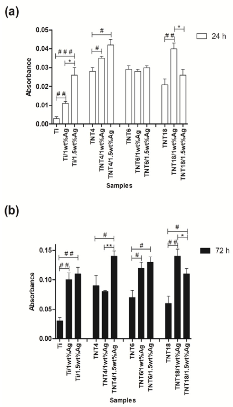
Figure 4.
Effect of TiO2 nanotube coatings enriched with the different concentrations of silver nanoparticles (1wt% and 1.5wt%; TNT/Ag) on the L929 cell adhesion (after 24 h; (a)) and proliferation (after 72 h; (b)) on the nanotube surface detected by the MTT assay. The absorbance values are expressed as means ± standard error mean (S.E.M.) of three experiments. Asterisk indicates significant differences between the cells incubated with the respective nanotubes coating doped by different concentrations of Ag (1% vs. 1.5%; * p < 0.05, ** p < 0.01). Hash mark indicates significant differences between the cells incubated with TiO2 nanotubes or Ti plates (TNT or Ti) in comparison to the cells incubated with respective TNT or Ti coatings doped by silver nanoparticles (TNT/Ag or Ti/Ag; # p < 0.05, ## p < 0.01, ### p < 0.001).

