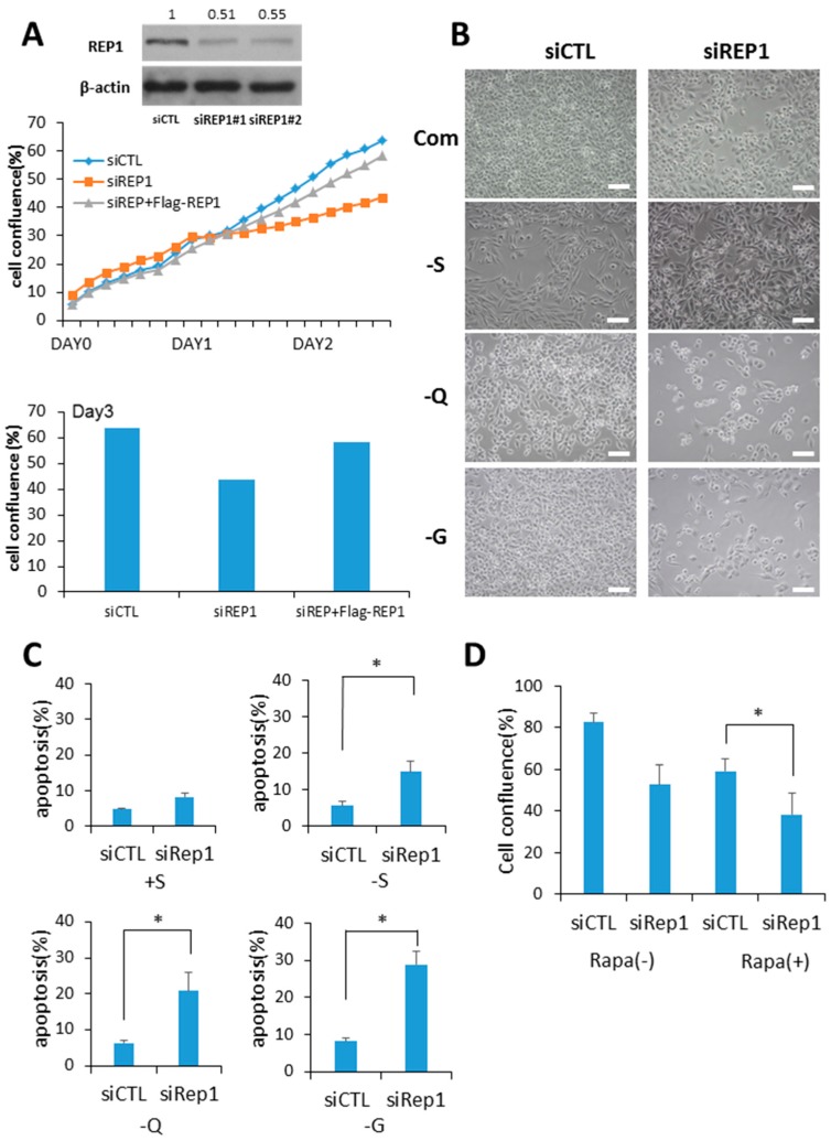
Figure 1.
Rab escort protein1 (REP1) depletion suppresses cell growth and survival. (A) MiaPaCa2 cells were transfected with control (CTL) and REP1 small interfering RNAs (siRNAs). After 48 h, immunoblotting was performed to analyze REP1 protein levels. Cells were treated with CTL and REP1 siRNAs and incubated for 24 h. Then, the cells were transfected with Flag-REP1 plasmid additionally and further incubated in the IncuCyteTM for monitoring cell proliferation. At the 72-h incubation time point, cell confluence levels were presented as a percentage using the IncuCyteTM analyzer. (B,C) MiaPaCa2 cells were transfected with control and REP1siRNAs, which replaced the following day with serum-, glucose-, or glutamine-free medium and then incubated for another 24 h.Cell morphology was observed by brightfield image. Scale bar: 50 μm (B). Cell death was assessed by using the Annexin V/propidium iodide (PI) assay (C). Error bars indicate mean +/− standard error for n = 3 independent experiments. (D) MiaPaCa2 cells were transfected with control (CTL) or REP1siRNAs, which replaced the following day with 1 µM rapamycin and then further incubated for monitoring cell confluence using IncuCyteTM. At 72 h time point, cell confluence levels were presented as percentage. Statistical significance was determined via t-test; * p < 0.05.

