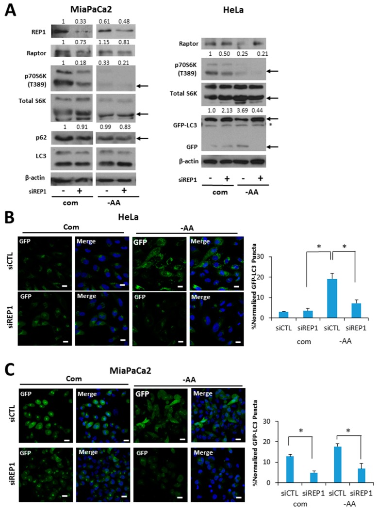
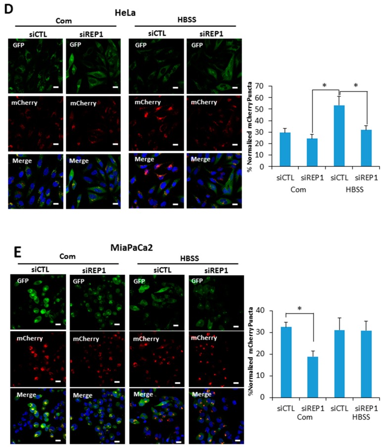
Figure 3.
REP1 depletion suppresses starvation-induced autophagy. (A) Hela and MiaPaCa2 cells stably expressing GFP-LC3 with a retroviral vector system were treated with CTL or REP1 siRNA and incubated for 48 h. Then, after 2 h of incubation in amino acid-deprived media, cells were harvested and immunoblotted for LC3, P62, and GFP. These results are representative of more than three independent experiments. GFP-LC3 puncta were analyzed by live imaging using fluorescence microcopy in (B) Hela and (C) MiaPaCa2 cells. Scale bar: 20 μm. Quantification data for GFP-LC3 puncta area are expressed as a percentage of the DAPI area within the cell. Hela and MiaPaCa2 cells stably expressing mCherry-GFP-LC3 with a retroviral vector system were treated with CTL or REP1 siRNA and incubated for 48 h. Then, after 2 h of incubation in Hank’s balanced saline solution (HBSS) media, mCherry puncta from mCherry-GFP-LC3 were imaged by fluorescence microcopy in (D) Hela and (E) MiaPaCa2 cells. Scale bar: 20 μm. Quantification data for mCherry puncta area derived from mCherry GFP-LC3 are expressed as a percentage of the DAPI area within the cell. Images shown are representative of at least three independent experiments. Statistical significance was determined via t-test; * p < 0.05.


