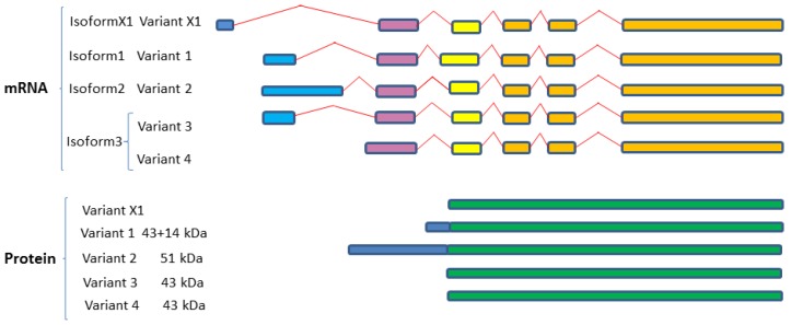
Figure 3.
Variant splicing and dicing of human SphK1 isoforms. Schematic diagram of SphK1 splice variants and protein isoforms based on mRNA and protein sequences acquired from GenBank. Colored boxes are representative of different RNA fragments and protein fragments, same color boxes are originated from the same origin of DNA sequences and are identical/similar sequences. Expression from four variant mRNA transcripts (variants 1–4) results in three SphK1 isoforms (isoforms 1–3). A predicted fifth human SphK1 splice variant (variant X1), based on sequence prediction methods, results in a predicted fourth isoform (isoform X1) (annotated using gene prediction software and further evidenced by mRNA and EST). SphK1 sequences were aligned using Clustal Omega (V1.2.4) multiple sequence alignment (Available online: http://www.ebi.ac.uk/Tools/msa/clustalo/).

