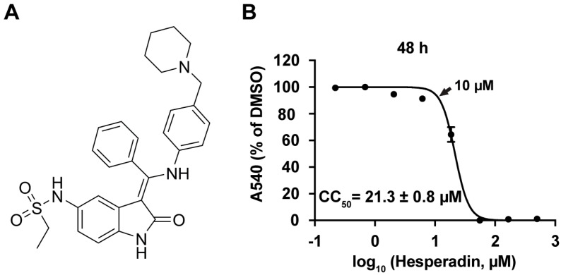
Figure 1.
(A) Chemical structure of hesperadin; (B) Cytotoxicity of hesperadin. Serial concentrations of hesperadin were added to Madin-Darby Canine Kidney (MDCK) cells and incubated for 48 h. The cell viability was determined by neutral red up-taken assay [24]. The CC50 was calculated from the best-fit dose response curves with variable slope in Prism 5. The arrow indicates the highest concentration used in Figure 2. The CC50 value represents the average of eight repeats ± standard deviation.

