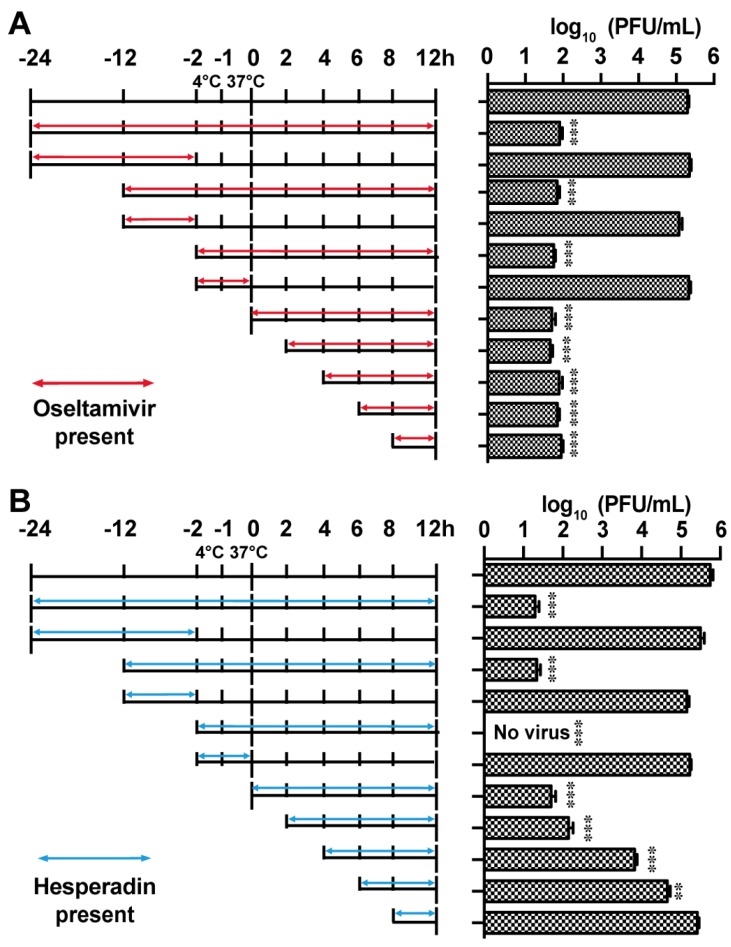
Figure 4.
Time-of-addition experiments. MDCK cells were infected with the A/WSN/33 (H1N1) at MOI of 0.01 at −2 h time point; viruses were first incubated at 4 °C for 1 h for attachment followed by 37 °C for 1 h for viral entry. At time point 0 h, cells were washed with PBS buffer and viruses were harvested at 12 h p.i. The titer of harvested virus was determined by plaque assay. Arrows indicate the period in which (A) 1 µM oseltamivir carboxylate or (B) 3 µM hesperidin was present. Asterisks indicate statistically significant difference in comparison with the DMSO control (student’s t-test, ** p < 0.01, *** p < 0.001). The results represent the average of two repeats ± standard deviation.

