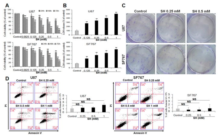
Figure 1.
SH inhibits cell proliferation through a caspase-independent pathway in U87 and SF767 cells. (A) Dose- and time-dependent effects of SH on cell viability. U87 and SF767 cells were treated with different concentrations of SH (0.0625, 0.125, 0.25, 0.5, 1 mM) for 24, 48 or 72 h, and cell viability was assessed via cell counting kit-8 (CCK-8) assays; (B) The cells were treated with 0.125, 0.25, 0.5, or 1 mM SH for 48 h, and LDH assays were performed; (C) U87 and SF767 cells viability upon the indicated concentrations of SH treatment was measured via colony formation assays; (D) Dose-dependent effects of SH on apoptosis. After treatment with the indicated concentrations of SH for 48 h, apoptosis was detected through Annexin V-FITC/PI staining and quantitative analysis of the percentage of Annexin V-positive and PI-negative cells compared with the control groups; (E) Extent of apoptosis, as observed by the levels of cleaved caspase-3 in U87 and SF767 cells, in the two cell lines treated with the indicated concentrations of SH for 48 h or at 1 mM for the indicated times; (F) Effects of the caspase-3 inhibitor Ac-DEVD-CHO and the pancaspase inhibitor Z-VAD-FMK on human glioblastoma cell death induced by SH. U87 and SF767 cells were preincubated with Z-VAD-FMK (50 µM) and Ac-DEVD-CHO (50 µM) for 1 h, followed by co-incubation with SH (1 mM) for 48 h. Each image is representative of n = 3 experiments. The results shown in E are one representative Western blot, n = 3, and β-actin served as the loading control. All data are shown as the mean ± SEM, n = 3. * p < 0.05, ** p < 0.01, versus the control. NS, not significant versus the control or SH treatment alone.


