Figure 3.
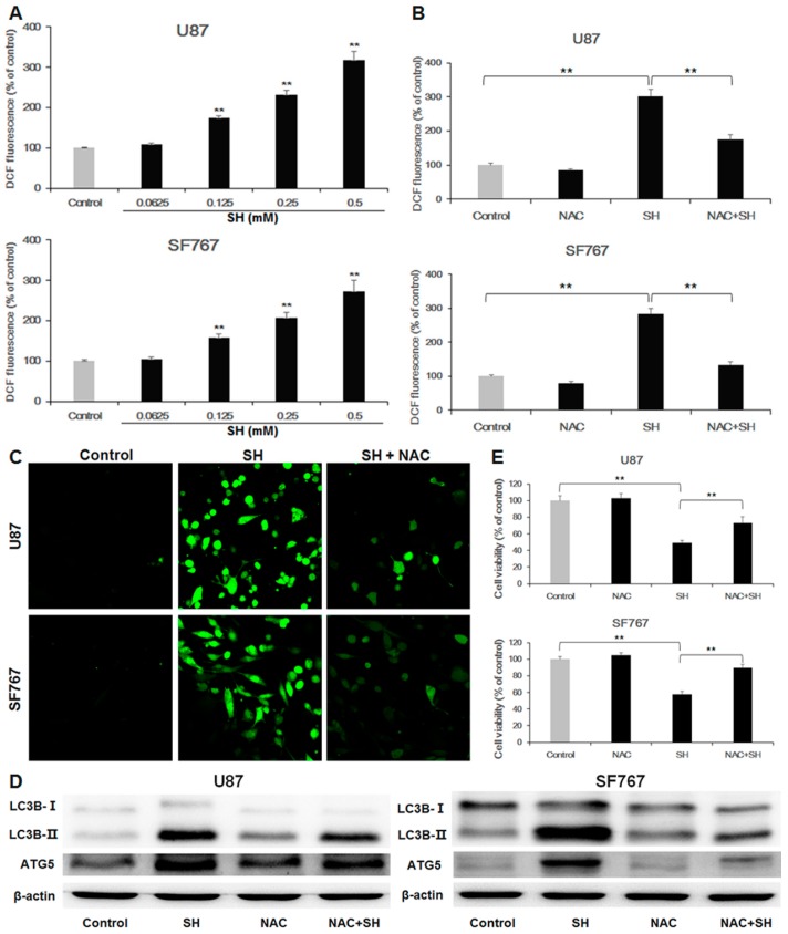
SH induces autophagy-mediated cell growth inhibition via ROS production (A) Dose-dependent effects of SH on intracellular ROS levels. U87 and SF767 cells were treated with different concentrations of SH for 24 h, and cellular ROS levels were detected using DCFH-DA probes, with the resulting fluorescence being read by a fluorescence microplate reader; (B) Effects of NAC on SH-mediated ROS generation. After pretreatment with the antioxidant NAC, followed by treatment with SH (0.5 mM) for 24 h, ROS levels were tested; (C) After pretreatment with the antioxidant NAC, followed by treatment with SH (0.5 mM) for 48 h, the ROS levels were imaged via confocal microscopy; (D) Effects of NAC on SH-mediated expression levels of LC3B-II and ATG5 in U87 and SF767 cells. (E) Effects of NAC on SH-mediated cell growth inhibition in U87 and SF767 cells. After pretreatment with the antioxidant NAC, followed by treatment with SH (0.5 mM) for 48 h, cell viability was measured via CCK-8 assays. Each image is representative of n = 3 experiments. The results shown in (D) are a representative Western blot, n = 3, and β-actin served as the loading control. All data are shown as the mean ± SEM, n = 3. ** p < 0.01, versus the control or SH treatment alone.

