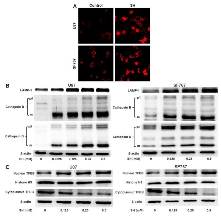
Figure 6.
SH facilitates lysosomal biogenesis in U87 and SF767 cells. (A) After 24 h of SH (0.5 mM) treatment, the cells were stained with LTR and imaged via confocal microscopy; (B) U87 and SF767 cells were treated with the indicated concentrations of SH for 24 h. The expression levels of lysosome markers (LAMP1, cathepsin B, cathepsin D) were observed via Western blotting analysis. p/i, precursor/intermediate; m, mature form of cathepsin B and cathepsin D; (C) Effects of SH on the nuclear translocation of TFEB. U87 and SF767 cells were treated with the indicated concentrations of SH for 24 h. The expression levels of TFEB in the cytosolic and nuclear fractions were detected through Western blotting analysis. Histone H3 and β-actin were used as loading controls for the nuclear and cytoplasmic fractions, respectively. Each image is representative of n = 3 experiments. All blots shown are representative of n = 3 experiments, with similar results.

