Figure 3.
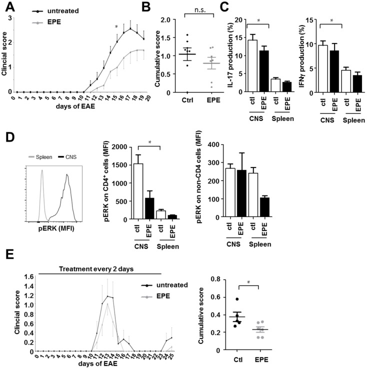
The inhibitory EPE peptide has only minor impact in EAE models in vivo. (A) Active EAE in C57BL/6 mice was induced by the injection of myelin oligodendrocyte glycoprotein (MOG)35–55/complete Freund's adjuvant (CFA) emulsion followed by pertussis toxin. EPE peptide was administered intraperitoneally (i.p.) two days before disease induction, and then every other day until day 18; (B) The cumulative score of all EAE animals treated with EPE and DMSO (as control) was assessed. On day 18 after EAE induction, leukocytes from the central nervous system (CNS) and splenocytes were stimulated ex vivo and stained for; (C) IL-17 and IFNγ; (D) pERK. Bar charts represent the mean percentage of nine mice and error bars show SEM (CD4+ cells: DMSO CNS vs. EPE CNS, p = 0.95 and DMSO Spleen vs. EPE Spleen, p = 0.09; non-CD4 cells DMSO Spleen vs. EPE Spleen, p = 0.06); (E) Active EAE was induced in female SJL mice via injection with murine PLP139–151. Intraperitoneal administration of DMSO as a solvent (n = 5) or EPE peptide (n = 6) started two days before immunization, and then every other day until day 20. EPE administration slightly reduced the EAE course; however, the cumulative score was significantly decreased by the treatment with EPE. Error bars show SEM. p-Values were obtained using unpaired student-t-test comparing two groups. * p < 0.05.

