Figure 3.

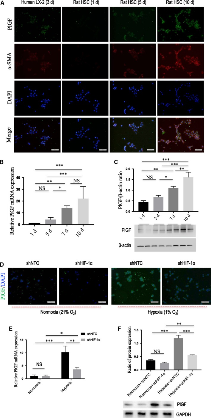
PlGF is overexpressed in activated hepatic stellate cells (HSCs) and its expression is induced by hypoxia dependent on HIF‐1α. (A) Immunofluorescent expression of PlGF (green) and α‐SMA (red) in LX‐2 human cell line and primary rat HSCs at different times in vitro activation; DAPI as blue nuclear counterstain. Scale bar = 100 μm for each picture. (B) The levels of PlGF mRNA expression in rat HSCs at different times in vitro activation were examined by quantitative RT‐PCR (n = 5). (C) The levels of PlGF protein expression in rat HSCs at different time points in vitro activation were examined by Western blot. (D) Immunofluorescent expression of PlGF (green) in rat HSCs; DAPI as blue nuclear counterstain. After infection with HIF‐1α shRNA (shHIF‐1α) or NTC shRNA (shNTC), cells were cultivated under hypoxic conditions (1% O2) or normoxia (21% O2) for 24 hrs. Scale bar = 100 μm for each picture. (E) The levels of PlGF mRNA expression in rat HSCs were measured by quantitative RT‐PCR. (F) Western blot analysis demonstrating effective silencing of HIF‐1α on the expression of PlGF in rat HSC, and GAPDH as loading control (n = 3).
