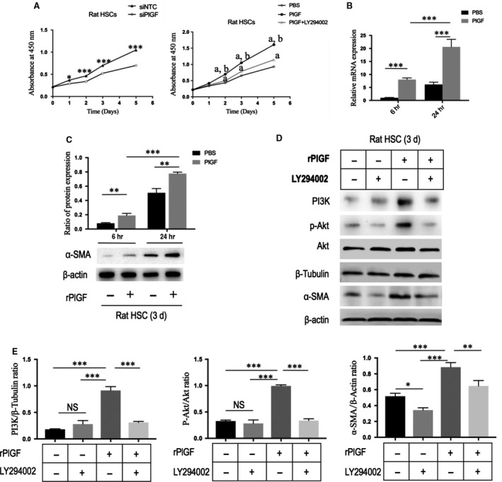
Figure 8.

PlGF knockdown by siRNA inhibits the proliferation and activation of hepatic stellate cells (HSCs) via the PI3K/Akt signalling pathway. (A) Measurement of cell proliferation of rat HSCs using CCK‐8 assay. Cells were transfected with PlGF siRNA or NTC siRNA; cells were treated with rPlGF administration (50 ng/ml) or co‐incubation with PI3K inhibitor LY294002 for 5 days. aP < 0.001 compared with mimics control (PBS), bP < 0.001 compared with PlGF+LY294002. (B) The mRNA levels of α‐SMA in rat HSCs with stimulation PlGF (50 ng/ml) for 6 or 24 hrs. (C) Representative Western blot of α‐SMA expression in rat HSC treated with rPlGF (50 ng/ml) or PBS for 6–24 hrs and quantification compared to β‐actin content. (D) Western blot for PI3K, phospho‐Akt (p‐Akt), Akt and α‐SMA in rat HSCs, and β‐tubulin or β‐actin as loading controls. Cells were treated with rPlGF administration (50 ng/mL) or co‐incubation with PI3K inhibitor LY294002 for 8 hrs. (E) The Western blot results of part (D) were quantified by densitometry.
