
Figure 5.
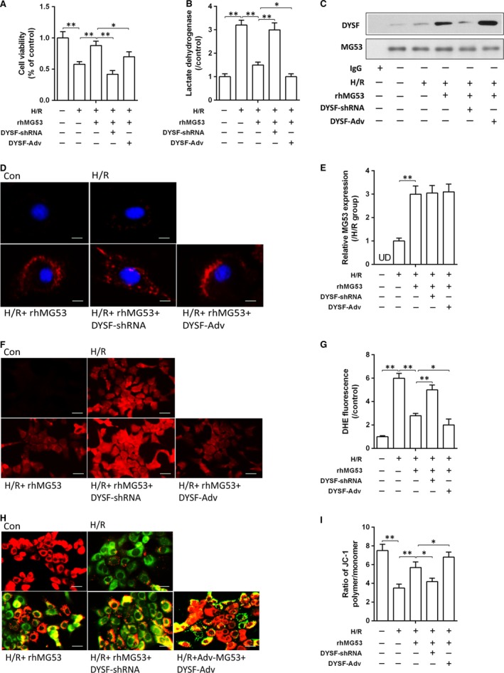
Down‐regulation of dysferlin reversed the protective effects of rhMG53 on hepatocyte H/R damage. (A and B) Cell proliferation was detected by CCK‐8 and cellular injury analysed by measuring LDH release in AML‐12 hepatocytes after H/R stimulation; (C) immunoprecipitation assay of dysferlin/MG53 of hepatocytes. (D) Immunofluorescence of MG53 protein expression on single hepatocyte using laser confocal microscopy (×1000); (E) density analysis of MG53 protein expression; (F and G) reactive oxygen species (ROS) levels assessed using fluorescence microscopy following the changes in DHE fluorescence (×400); (H and I) mitochondrial membrane potential (MMP) loss measured using a JC‐1 mitochondrial membrane potential assay method (×400). Date are mean ± SEM, and each experiment was performed at least independently in triplicate, *P < 0.05, **P < 0.01. H/R, hypoxia/reoxygenation; rhMG53, recombinant human MG53 protein; DYSF, dysferlin; UD, undetectable.