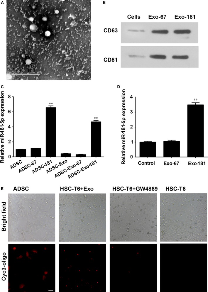
Figure 2.

Exosome‐mediated miR‐181‐5p communication between adipose‐derived mesenchymal stem cells (ADSCs) and HST‐T6 cells. (A) Exosomes extracted from ADSCs were identified by TEM. Magnification: ×150,000. Scale bar: 500 nm (B): Western blot for CD63 and CD81 expression in ADSC‐derived exosomes. Real‐time PCR detection of miR‐181‐5p expression in ADSCs, ADSC‐derived exosomes (C) and exosome‐treated HST‐T6 cells (D). (E) Confocal images of ADSC‐181 stained with cyc3‐oligo. Transfer of fluorescent exosomes from ADSC‐181 is apparent in HST‐T6 cell membranes and cytoplasm. Data are presented as means ± SE. (**P < 0.01, n = 3). ADSC‐181: miR‐181‐5p‐transfected ADSCs; HST‐T6 + Exo: ADSC‐derived exosomes transfer of miRNA from the ADSCs to the target cells; HST‐T6 + GW4869:GW4869 (10 M for 48 hrs) exosomal inhibitor, was confirmed to inhibit the elevated expression of miR‐181‐5p in HST‐T6 cells when cocultured with miR‐181‐5p‐ADSCs. HST‐T6: Brightfield and immunofluorescence images showing HST‐T6 cells without cyc3‐oligo transfection. Scale bar = 100 μm.
