
Figure 4.
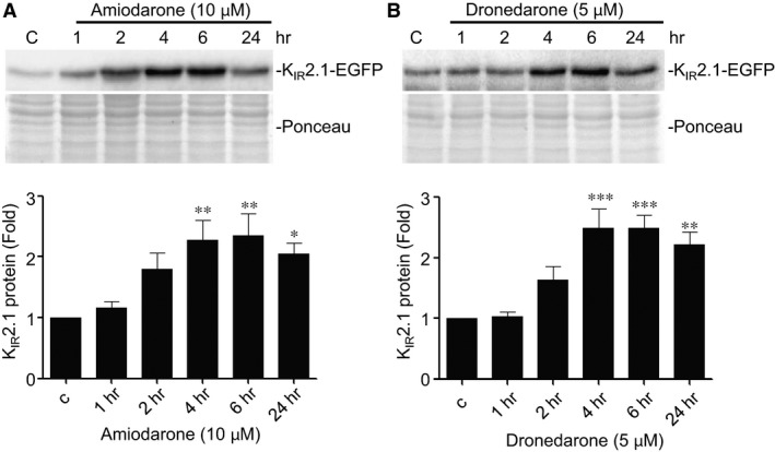
Amiodarone and dronedarone induce time‐dependent increases in KIR2.1‐GFP expression. Western blot analysis of KIR2.1‐GFP expression in HK‐KWGF cells treated for 1, 2, 4, 6 and 24 hrs with 10 μM amiodarone or 5 μM dronedarone. C indicates control (untreated) cells. Ponceau staining is used as loading control. Averaged data from eight (amiodarone) and ten (dronedarone) independent experiments, respectively, are shown in bar graphs in the lower part of both panels. *P < 0.05; **P < 0.01; ***P < 0.001.