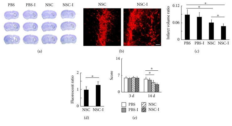
Figure 3.
The inhibition of striatal neuronal activity enhanced the survival and migration of transplanted NSC and further reduced brain infarct volume. (a) Cresyl violet staining of the brain infarction at 14 days after tMCAO. (b) The fluorescence of NSC-RFP in the striatum at 14 days after tMCAO, bar = 100 μm. (c) The ratio of infarct volume over the contralateral hemisphere volume (PBS = 8.6 ± 1.4%, PBS-I = 8.1 ± 1.6%, NSC = 6.3 ± 0.8%, and NSC-I = 4.5 ± 0.7%, n = 7). (d) The quantification of fluorescence signal from the transplanted NSCs (NSC = 1.00 ± 0.17, NSC-I = 1.29 ± 0.18, n = 6). (e) The behavioral outcome evaluated by NSS method at 3 days (PBS = 5.9 ± 0.6, PBS-I = 5.7 ± 0.4, NSC = 6.1 ± 0.4, and NSC-I = 5.9 ± 0.5, n = 7) and 14 days (PBS = 5.5 ± 0.9, PBS-I = 5.1 ± 0.8, NSC = 4.0 ± 0.9, and NSC-I = 3.3 ± 1.0, n = 7) after tMCAO. ∗ represents p < 0.05.

