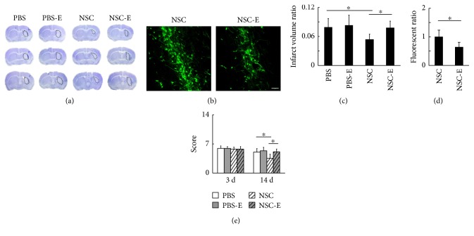
Figure 4.
The excitation of striatal neurons after NSC transplantation reversed the beneficial effects of NSC in reducing brain infarction. (a) The brain infarction at 14 days after tMCAO. (b) The fluorescence from NSC-GFP in the striatum at 14 days after tMCAO, bar = 100 μm. (c) The ratio of infarct volume over the contralateral hemisphere volume (PBS = 7.9 ± 1.8%, PBS-E = 8.3 ± 2.1%, NSC = 5.4 ± 1.1%, and NSC-E = 7.8 ± 1.4%, n = 7). (d) The quantification of fluorescence signal from the transplanted NSCs (NSC = 1.00 ± 0.23, NSC-E = 0.64 ± 0.16, n = 6) (e) The behavioral outcome evaluated by NSS method at 3 days (PBS = 5.7 ± 0.6, PBS-E = 5.7 ± 0.4, NSC = 5.6 ± 0.5, and NSC-E = 5.6 ± 0.6, n = 7) and 14 days (PBS = 4.8 ± 0.8, PBS-E = 5.1 ± 0.8, NSC = 3.4 ± 1.0, and NSC-E = 4.9 ± 0.7, n = 11) after tMCAO. ∗ represents p < 0.05.

