. 2017 May 24;8(8):5585–5590. doi: 10.1039/c7sc00260b

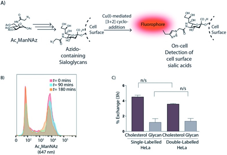
Fig. 4. (A) Schematic representation of cell surface glycan labelling. (B) Flow cytometry overlay histograms upon different times show no enhancement of glycolipid exchange between jurkat cells after coiled-coil formation; jurkat cells were treated with Ac4ManNAz and CPE co-cultured with untreated cells with CPK and labelled with CuAAC. (C) ΔMFI [ΔMFI = (MFInegative (180 min) – MFInegative (0 min))/MFIpositive (0 min)] expression of exchange after 3 h between sterol and glycan in single- and double-labelled co-culture experiments; data show lipid exchange independently of the glycan exchange. Data are represented as mean ± SD. Error bars SD; n/s p > 0.05; unpaired t-tests.

Fig. 4