Figure 1.
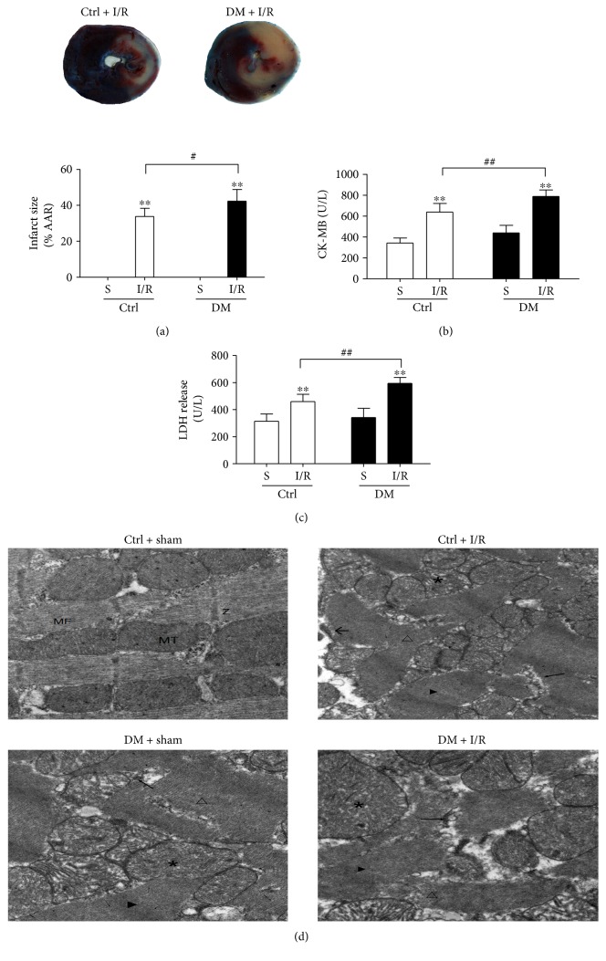
Diabetes aggravated the degree of MI/R injury in rats. The infarct size was detected by TTC staining (a). The levels of CK-MB and LDH release were determined by enzyme activity assay kits ((b) and (c)). The ultrastructural changes of rat hearts were detected by TEM (d): normal myofibrils (MF); normal mitochondria (MT); normal Z-lines (Z); disorganized myofibrils (△); swollen mitochondria (∗); expanded sarcoplasmic reticulum (←); disappearance of the Z-line (▶); and dissolved muscle cell membrane (←). Data are expressed as the mean ± SD. n = 6. ∗∗P < 0.01 versus sham; #P < 0.05 and ##P < 0.01 versus Ctrl + IR.

