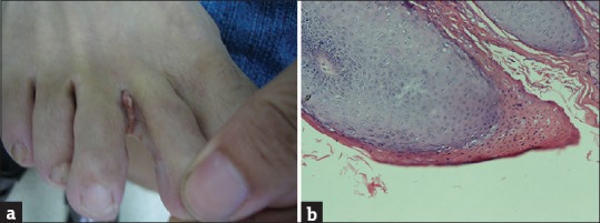
Figure 1.

(a) Spiky cauliflower-shaped verrucous lesions in case 1. (b) Parakeratosis and hyperkeratosis in the epidermis, thickened granular layer, and acanthosis. In the granular layer, giant cells with vacuolated cytoplasm (koilocytotic cells) were present, indicating human papillomavirus infection in case 1(haematoxylin and eosin staining; original magnification, ×100)
