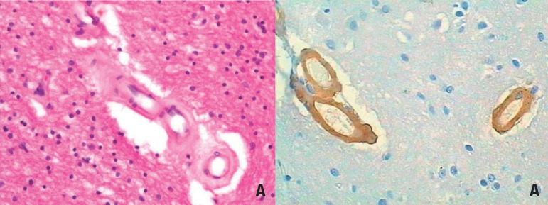
Figure 1.
Example of small vessel disease. [A] Subcortical white matter exhibiting hyaline arteriolosclerosis. Note how the arterial wall is thickened by hyaline material. HE. 200x. [B] Cortex exhibiting cerebral amyloid angiopathy. Amyloid-β peptide infiltrates the vessel wall. 200x. Immunohistochemistry against amyloid-β.

